Left Hip Muscles Anatomy / Iliopsoas Wikipedia. Ligaments, tendons, and muscles play an important role in the function of the hip. Human muscle system, the muscles of the human body that work the skeletal system, that are under voluntary control, and that are concerned with movement, posture, and balance. These ligaments reinforce and stabilize the hip joint(6). The hip's essential muscles are the sartorius, rectus femoris, gluteus minimus and medius, iliopsoas, adductors, and hamstrings. Most modern anatomists define 17 of these muscles, although some additional muscles may sometimes be considered.
Functionally, the hip joint enjoys a very high range of motion. Any injury or disease of the hip will adversely affect the joint's range of motion and ability to bear weight.</p> Anatomography, 2013 the hip flexors can be found connecting the top of the femur, which is the largest bone in the body, to the. Anatomy of the hip muscles. These are gracilis, pectineus, adductor longus, adductor brevis, adductor magnus, and adductor minimus muscles.
Nlrfkayqknn0mm from mk0hippainhelp9h8quy.kinstacdn.com There are four gluteal muscles, located at the posterior side of the hip bone: The posterior muscle group is made up of the muscles that extend (straighten) the thigh at the hip. There are 3 main layers of hip abductor muscles: Related posts of muscles of the lower back and hip diagram muscle anatomy app. Either left pelvis rotates downward or right pelvis rotates upward; The thigh bone or femur and the pelvis join to form the hip joint. It proceeds across the iliopubic eminence through the muscular lacuna to its insertion on the lesser trochanter of the femur. The anatomy of the hip abductors has not been comprehensively examined, yet is important to understanding function and pathology in the gluteal region.
The movements that can be carried out at the hip joint are listed below, along with the principle muscles responsible for each action:
The iliofemoral, pubofemoral, and ischiofemoral ligaments represent the thickenings of the joint capsule. Adductor muscles on the inside of your thigh. Muscle anatomy app 12 photos of the muscle anatomy app best muscle anatomy app, best muscle anatomy app for android, free muscle anatomy app, human muscle anatomy app, muscle anatomy app ipad, human muscles, best muscle anatomy app, best muscle anatomy app for android, free muscle anatomy app, human muscle. Broadly considered, human muscle—like the muscles of all vertebrates—is often divided into striated muscle, smooth muscle, and cardiac muscle. Some of the other muscles in the hip are: It joins the psoas major to form the iliopsoas. The movements that can be carried out at the hip joint are listed below, along with the principle muscles responsible for each action: The psoas muscles are part of the hip flexor muscles. Iliopsoas muscle, a hip flexor muscle that attaches to the upper thigh bone. Semimembranosus, semitendinosus and biceps femoris (the hamstrings) One at the left hip, and one at the right hip. Rectus femoris muscle, one of. The hip's essential muscles are the sartorius, rectus femoris, gluteus minimus and medius, iliopsoas, adductors, and hamstrings.
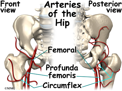
Anatomy of the hip muscles. Blood vessels and nerves of the hip It is also referred to as a ball and socket joint and is surrounded by muscles, ligaments, and tendons. Rectus femoris muscle, one of. It joins the psoas major to form the iliopsoas.
Hip Anatomy Orthogate from www.eorthopod.com The iliacus arises from the iliac fossa on the interior side of the hip bone, and also from the region of the anterior inferior iliac spine (aiis). Hip pain on the outside of your hip, upper thigh or outer buttock is usually caused by problems with muscles, ligaments, tendons and other soft tissues that surround your hip joint. Posterior view of gluteus maximus and gluteus medius in human anatomy, the muscles of the hip joint are those muscles that cause movement in the hip. The femur may also rotate around its axis about 90 degrees at the hip. Tightness in this muscle can compress the sciatic nerve. Functionally, the hip joint enjoys a very high range of motion. Semimembranosus, semitendinosus and biceps femoris (the hamstrings) Anatomy of the hip muscles.
Functionally, the hip joint enjoys a very high range of motion.
The six hip adductor muscles are all located in the adductor or medial compartment of the thigh and all mainly adduct the thigh at the hip joint. These ligaments reinforce and stabilize the hip joint(6). Ligaments are soft tissue structures that connect bones to bones.a joint capsule is a watertight sac that surrounds a joint.in the hip, the joint capsule is formed by a group of three strong ligaments that connect the femoral head to the acetabulum. Anatomy of the hip muscles. These are gracilis, pectineus, adductor longus, adductor brevis, adductor magnus, and adductor minimus muscles. They begin under the gluteus maximus behind the hipbone and attach to the tibia at the knee. The psoas muscles are part of the hip flexor muscles. The bones of the hip include the femur, the ilium, the ischium, and the pubis. Functionally, the hip joint enjoys a very high range of motion. Broadly considered, human muscle—like the muscles of all vertebrates—is often divided into striated muscle, smooth muscle, and cardiac muscle. The hip joint is the articulation of the pelvis with the femur, which connects the axial skeleton with the lower extremity. Tightness in this muscle can compress the sciatic nerve. Some of the organs include the descending.
Iliopsoas muscle, a hip flexor muscle that attaches to the upper thigh bone. The sartorius muscle is a distinctively long and thin muscle that crosses the thigh diagonally. Either left pelvis rotates downward or right pelvis rotates upward; The movements that can be carried out at the hip joint are listed below, along with the principle muscles responsible for each action: The iliofemoral, pubofemoral, and ischiofemoral ligaments represent the thickenings of the joint capsule.
Muscles Of The Hips And Thighs Human Anatomy And Physiology Lab Bsb 141 from s3-us-west-2.amazonaws.com The iliofemoral, pubofemoral, and ischiofemoral ligaments represent the thickenings of the joint capsule. The hip's essential muscles are the sartorius, rectus femoris, gluteus minimus and medius, iliopsoas, adductors, and hamstrings. The psoas muscles are part of the hip flexor muscles. Anatomy of the hip muscles. These muscles include the gluteus maximus muscle (the largest muscle in the body) and the hamstrings group, which consists of the biceps femoris, semimembranosus, and semitendinosus muscles. Since the upper part of the pelvic bone lies within the abdominal area, any organs in this region can be possible causes of pain on left side above hip. These ligaments reinforce and stabilize the hip joint(6). Hip flexor connects the top femur to the lower back as well as the hips and the groin
Any injury or disease of the hip will adversely affect the joint's range of motion and ability to bear weight.</p>
It proceeds across the iliopubic eminence through the muscular lacuna to its insertion on the lesser trochanter of the femur. The thigh bone or femur and the pelvis join to form the hip joint. Understanding the possible causes of hip flexor pain has a number of benefits. The ball is the femoral head, and the socket is the acetabulum. Semimembranosus, semitendinosus and biceps femoris (the hamstrings) Since the upper part of the pelvic bone lies within the abdominal area, any organs in this region can be possible causes of pain on left side above hip. The posterior muscle group is made up of the muscles that extend (straighten) the thigh at the hip. One at the left hip, and one at the right hip. Anatomography, 2013 the hip flexors can be found connecting the top of the femur, which is the largest bone in the body, to the. Tightness in this muscle can compress the sciatic nerve. The anatomy of the hip abductors has not been comprehensively examined, yet is important to understanding function and pathology in the gluteal region. The anatomy of the fascia lata and iliotibial tract; Gluteus maximus, gluteus medius, gluteus minimus, and tensor fasciae latae.
Share :
Post a Comment
for "Left Hip Muscles Anatomy / Iliopsoas Wikipedia"


Post a Comment for "Left Hip Muscles Anatomy / Iliopsoas Wikipedia"